Description
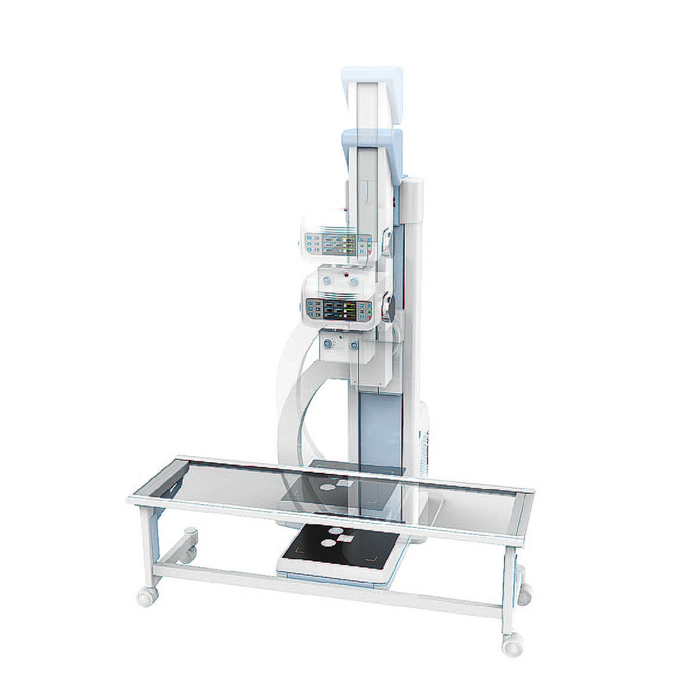
A C-Arm machine is an advanced medical imaging device based on X-ray technology. They are primarily used for fluoroscopy capabilities, although they have radiography capability too. C-Arm is called so, due to its C-shaped arm, which is used to connect the x-ray source on one end and the detector on the other.
C-Arm machines are widely used during orthopedic, urology, gastroenterology surgeries, other complicated surgical, pain management and emergency procedures, cardiac and angiography studies and in therapeutic studies including stents or needle placements. The Fluoroscopy technology enables high-resolution X-ray images provided in real time, so that the surgeons can do more accurate procedures with better outcomes for the patient. C-Arm systems are also mobile and provide a great deal of flexibility to the surgeon to take images of whole body of the patient from different angles as required.
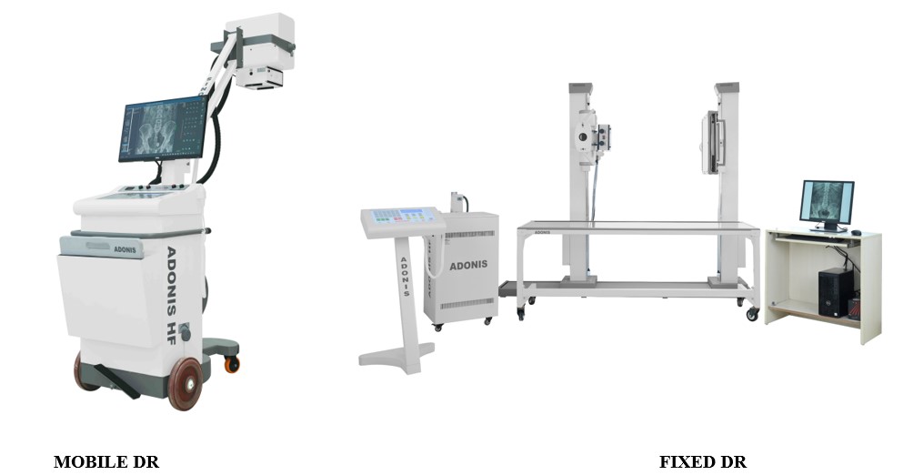
How does a mobile C-arm machine work? What are its key components?
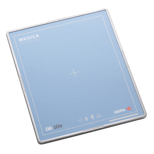
A mobile C-arm, comprises of a X-Ray generator, an image intensifier or flat-panel detector and workstation, which has the controls and settings. The C-shaped arm connecting the generator and detector, allows movement horizontally, vertically and around the swivel axes, so that X-ray images of the patient are produced from many angles. The generator emits X-rays that penetrate the patient’s body which is placed in between. The image intensifier or detector converts the X-rays into a visible image displayed on the C-arm monitor. Physician can check anatomical details such as bones, fractures in the bone under consideration vs the position of implants and instruments in real-time, while doing the procedure, without causing damage to other parts.